[fullwidth background_color=”#ebebeb” background_image=”” background_parallax=”none” enable_mobile=”no” parallax_speed=”0.3″ background_repeat=”no-repeat” background_position=”left top” video_url=”” video_aspect_ratio=”16:9″ video_webm=”” video_mp4=”” video_ogv=”” video_preview_image=”” overlay_color=”” overlay_opacity=”0.5″ video_mute=”yes” video_loop=”yes” fade=”no” border_size=”0px” border_color=”” border_style=”solid” padding_top=”0″ padding_bottom=”0″ padding_left=”” padding_right=”” hundred_percent=”yes” equal_height_columns=”no” hide_on_mobile=”no” menu_anchor=”” class=”” id=””][imageframe lightbox=”no” lightbox_image=”” style_type=”none” hover_type=”none” bordercolor=”” bordersize=”0px” borderradius=”0″ stylecolor=”” align=”center” link=”” linktarget=”_self” animation_type=”0″ animation_direction=”down” animation_speed=”0.1″ hide_on_mobile=”no” class=”” id=””]  [/imageframe][/fullwidth][fullwidth background_color=”#ffffff” background_image=”” background_parallax=”none” enable_mobile=”no” parallax_speed=”0.3″ background_repeat=”no-repeat” background_position=”left top” video_url=”” video_aspect_ratio=”16:9″ video_webm=”” video_mp4=”” video_ogv=”” video_preview_image=”” overlay_color=”” overlay_opacity=”0.5″ video_mute=”yes” video_loop=”yes” fade=”no” border_size=”0px” border_color=”” border_style=”solid” padding_top=”40″ padding_bottom=”40″ padding_left=”” padding_right=”” hundred_percent=”no” equal_height_columns=”no” hide_on_mobile=”no” menu_anchor=”” class=”” id=””][two_third last=”no” spacing=”yes” center_content=”no” hide_on_mobile=”no” background_color=”” background_image=”” background_repeat=”no-repeat” background_position=”left top” border_position=”all” border_size=”0px” border_color=”” border_style=”” padding=”” margin_top=”” margin_bottom=”” animation_type=”” animation_direction=”” animation_speed=”0.1″ class=”” id=””][fusion_text]
Hydrocephalus
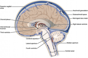
Hydrocephalus refers to enlargement of the fluid containing cavities within the brain due to an abnormal build up of cerebrospinal fluid (CSF). Normally cerebrospinal fluid is made in the central cavities of the brain known as ventricles.  From here, the fluid travels via small ducts and openings to reach the outer surface of the brain and spinal cord where the fluid is reabsorbed. This creates an inside to outside flow. In an adult about 450ml of cerebrospinal fluid is made in a day and the same amount is reabsorbed. Disturbance in this delicate balance can be created by blockage of the fluid pathways, over-production or under-absorption of the fluid.
The commonest symptoms of impaired fluid circulation are headache, nausea, vomiting, blurring of vision and in acute circumstances decreased conscious state that may possibly lead to death. In chronic circumstances the symptoms can be slow in onset and include memory disturbance, incontinence, unsteady gait and cognitive decline.
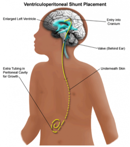
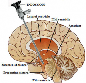
Treatment of hydrocephalus depends on the cause and symptoms. Two of the most common ways of treatment are the insertion of a shunt system, which diverts CSF, or the performance of an endoscopic third ventriculostomy, which creates a new, alternate flow pathway within the brain.
A shunt in its simplest form, is a tube that connects the cavities of the brain to a body cavity that has the potential to absorb the fluid. Most commonly the abdominal cavity is used for the diversion. The tube has a valve that controls the flow of the fluid from the brain cavity to the abdominal cavity. Most modern valves have a programmable function, whereby your surgeon can alter the relative flow of the fluid by using a remote control. Fine adjustments can be made without needing to change the valve itself.
 Endoscopic third ventriculostomy refers to creating an alternate pathway from the central brain cavity (ventricle) for fluid to reach the outer surface of the brain for absorption. A camera (endoscope) is inserted into the ventricle and a perforation is made under direct vision to create the pathway.
Surgeons at Neurosurgery Tasmania are trained and experienced in all forms of fluid diversion procedures and will be able to assess your situation and recommend the most appropriate modality of treatment.[/fusion_text][/two_third][one_third last=”yes” spacing=”yes” center_content=”no” hide_on_mobile=”no” background_color=”#f7f7f7″ background_image=”” background_repeat=”no-repeat” background_position=”left top” border_position=”all” border_size=”0px” border_color=”” border_style=”solid” padding=”20px” margin_top=”” margin_bottom=”” animation_type=”0″ animation_direction=”down” animation_speed=”0.1″ class=”” id=””][fusion_widget_area name=”avada-custom-sidebar-otherneurosurgicalconditions” background_color=”#f7f7f7″ padding=”10px” class=”” id=””][/fusion_widget_area][/one_third][/fullwidth]


