[fullwidth background_color=”#ebebeb” background_image=”” background_parallax=”none” enable_mobile=”no” parallax_speed=”0.3″ background_repeat=”no-repeat” background_position=”left top” video_url=”” video_aspect_ratio=”16:9″ video_webm=”” video_mp4=”” video_ogv=”” video_preview_image=”” overlay_color=”” overlay_opacity=”0.5″ video_mute=”yes” video_loop=”yes” fade=”no” border_size=”0px” border_color=”” border_style=”solid” padding_top=”0″ padding_bottom=”0″ padding_left=”” padding_right=”” hundred_percent=”yes” equal_height_columns=”no” hide_on_mobile=”no” menu_anchor=”” class=”” id=””][imageframe lightbox=”no” lightbox_image=”” style_type=”none” hover_type=”none” bordercolor=”” bordersize=”0px” borderradius=”0″ stylecolor=”” align=”center” link=”” linktarget=”_self” animation_type=”0″ animation_direction=”down” animation_speed=”0.1″ hide_on_mobile=”no” class=”” id=””]  [/imageframe][/fullwidth][fullwidth background_color=”#ffffff” background_image=”” background_parallax=”none” enable_mobile=”no” parallax_speed=”0.3″ background_repeat=”no-repeat” background_position=”left top” video_url=”” video_aspect_ratio=”16:9″ video_webm=”” video_mp4=”” video_ogv=”” video_preview_image=”” overlay_color=”” overlay_opacity=”0.5″ video_mute=”yes” video_loop=”yes” fade=”no” border_size=”0px” border_color=”” border_style=”solid” padding_top=”40″ padding_bottom=”40″ padding_left=”” padding_right=”” hundred_percent=”no” equal_height_columns=”no” hide_on_mobile=”no” menu_anchor=”” class=”” id=””][two_third last=”no” spacing=”yes” center_content=”no” hide_on_mobile=”no” background_color=”” background_image=”” background_repeat=”no-repeat” background_position=”left top” border_position=”all” border_size=”0px” border_color=”” border_style=”” padding=”” margin_top=”” margin_bottom=”” animation_type=”” animation_direction=”” animation_speed=”0.1″ class=”” id=””][fusion_text]
Acoustic Neuroma
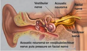
An acoustic neuroma (actually a misnomer, as the tumour typically arises on the vestibular component of the 8th cranial nerve, and may be more properly termed a vestibular Schwannoma), is a benign slow growing tumour, which initially appears within the internal auditory canal (the bony channel through which the 7th and 8th cranial nerves pass), and grows slowly into the cerebellopontine angle (the space between the inner surface of the skull and the cerebellum and brainstem).
 Acoustic neuromas may be present for many years before beginning to produce symptoms. This is possible because of their slow rate of growth, and the gradual adaption of the neural structures to the evolving pressure and displacement.
Typical early symptoms include gradual unilateral hearing loss (often initially high tone, and easily confused with other causes, such as noise related hearing loss, or hearing decline associated with aging).
Tinnitus (ringing in the ear) may also occur, and in some patients a feeling of disequilibrium or imbalance is prominent. Some patients report a feeling of fullness in the affected ear. Hearing loss is usually gradual, but on occasions abrupt decline in hearing may occur. As the tumour enlarges, it may compress and displace increasingly vital structures and, untreated, a fatal outcome due to brainstem compression would be possible.
With the increasing use of MRI scanning, the number of acoustic neuromas being diagnosed has increased, some as incidental findings when other pathologies are being looked for.
There are essentially three management options, and the most suitable will depend on the balance between a number of factors, including tumour size, its location, patient age, physical health, current symptoms and patient preference.
Observation, with serial MRI imaging
Small tumours, or non-compressive tumours in the elderly, may be monitored with annual MRI scans. Growth rates are usually slow, and sometimes imperceptibly so, and studies have shown that the most reliable way to preserve residual hearing is conservative management for as long as is reasonable.
Radiosurgery
Radiosurgery is a focused form of radiation treatment. It has been used for some years for small and small medium sized acoustic neuromas, and is reported to be successful in arresting growth in a large percentage of treated tumours.
The end point for successful treatment is markedly different for radiosurgery, as against microsurgery, and is closer to conservative management (no growth is considered success, but given the slow growth rate of many tumours, may be difficult to assess as differing from the natural history of the tumour, without a very extended period of follow-up). A relatively small number of treated tumours shrink, but very few, if any, disappear.
Radiosurgery may be complicated by delayed nerve injury, by higher complication rates if surgery is required due to tumour progression, and by a small chance of malignant change in the treated tumour.
It does, however, provide a viable alternative treatment choice, particularly if surgery is contraindicated by age or other medical conditions.
Microsurgery
Microsurgical tumour debulking, or, more commonly tumour excision, is performed by an open microscopically assisted operation.
There are a number of different approaches possible, but often a particular unit will specialise in one or two approaches. The retromastoid approach (through a small craniotomy located behind the affected ear) is the most versatile, and can be employed for all sizes of tumour. It is this approach that is employed by the team at Neurosurgery Tasmania. With this approach, hearing preservation is possible (but is unfortunately unpredictable), whilst with the translabyrinthine approach hearing is always lost. Similarly, there is a high rate of preserved facial nerve function, and the opportunity to perform nerve reconstruction exists if continuity cannot be maintained (a very rare circumstance).
Successful surgery will achieve a complete removal, and in most cases this is curative.
Long-term Management
All patients are likely to require long-term follow-up with serial MRI scans, generally annually. In the case of microsurgery , this is usually for 5 years after curative tumour resection. For radiosurgery it is not clear at what point surveillance can be ceased.[/fusion_text][/two_third][one_third last=”yes” spacing=”yes” center_content=”no” hide_on_mobile=”no” background_color=”#f7f7f7″ background_image=”” background_repeat=”no-repeat” background_position=”left top” border_position=”all” border_size=”0px” border_color=”” border_style=”solid” padding=”20px” margin_top=”” margin_bottom=”” animation_type=”0″ animation_direction=”down” animation_speed=”0.1″ class=”” id=””][fusion_widget_area name=”avada-custom-sidebar-cerebraltumours” background_color=”#f7f7f7″ padding=”10px” class=”” id=””][/fusion_widget_area][/one_third][/fullwidth]


