[fullwidth background_color=”#ebebeb” background_image=”” background_parallax=”none” enable_mobile=”no” parallax_speed=”0.3″ background_repeat=”no-repeat” background_position=”left top” video_url=”” video_aspect_ratio=”16:9″ video_webm=”” video_mp4=”” video_ogv=”” video_preview_image=”” overlay_color=”” overlay_opacity=”0.5″ video_mute=”yes” video_loop=”yes” fade=”no” border_size=”0px” border_color=”” border_style=”solid” padding_top=”0″ padding_bottom=”0″ padding_left=”” padding_right=”” hundred_percent=”yes” equal_height_columns=”no” hide_on_mobile=”no” menu_anchor=”” class=”” id=””][imageframe lightbox=”no” lightbox_image=”” style_type=”none” hover_type=”none” bordercolor=”” bordersize=”0px” borderradius=”0″ stylecolor=”” align=”center” link=”” linktarget=”_self” animation_type=”0″ animation_direction=”down” animation_speed=”0.1″ hide_on_mobile=”no” class=”” id=””] 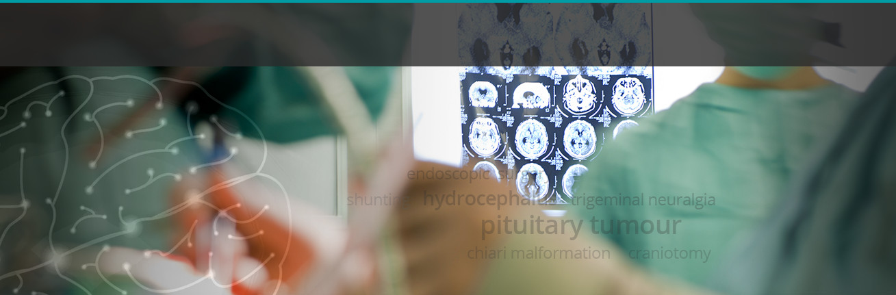 [/imageframe][/fullwidth][fullwidth background_color=”#ffffff” background_image=”” background_parallax=”none” enable_mobile=”no” parallax_speed=”0.3″ background_repeat=”no-repeat” background_position=”left top” video_url=”” video_aspect_ratio=”16:9″ video_webm=”” video_mp4=”” video_ogv=”” video_preview_image=”” overlay_color=”” overlay_opacity=”0.5″ video_mute=”yes” video_loop=”yes” fade=”no” border_size=”0px” border_color=”” border_style=”solid” padding_top=”40″ padding_bottom=”40″ padding_left=”” padding_right=”” hundred_percent=”no” equal_height_columns=”no” hide_on_mobile=”no” menu_anchor=”” class=”” id=””][two_third last=”no” spacing=”yes” center_content=”no” hide_on_mobile=”no” background_color=”” background_image=”” background_repeat=”no-repeat” background_position=”left top” border_position=”all” border_size=”0px” border_color=”” border_style=”” padding=”” margin_top=”” margin_bottom=”” animation_type=”” animation_direction=”” animation_speed=”0.1″ class=”” id=””][fusion_text]
Non-Communicating Hydrocephalus
Non-communicating hydrocephalus is due to blockage of the fluid in its course from the production site (central cavities) to the absorption site (the surface of the brain and spinal cord).
This can occur due to obstruction of the ducts or cavities by blood clots, tumours, infection related membranes or developmental anomalies. Often there will be asymmetric enlargement of the cavities with distortion of the brain that leads to symptom development.
Patients often develop acute symptoms that may require to urgent surgery to remedy the situation. Surgical technique can vary depending on the situation and the underlying pathology. In general, either the blockage is removed or the fluid is given alternate pathways or diverted to other cavities in the body to keep the intracranial volume constant.
Surgeons from Neurosurgery Tasmania are experienced in all forms of fluid diversion surgeries and use various techniques including using an endoscope to visualise the cavities and make alternate pathways using modern shunting techniques.[/fusion_text][/two_third][one_third last=”yes” spacing=”yes” center_content=”no” hide_on_mobile=”no” background_color=”#f7f7f7″ background_image=”” background_repeat=”no-repeat” background_position=”left top” border_position=”all” border_size=”0px” border_color=”” border_style=”solid” padding=”20px” margin_top=”” margin_bottom=”” animation_type=”0″ animation_direction=”down” animation_speed=”0.1″ class=”” id=””][fusion_widget_area name=”avada-custom-sidebar-otherneurosurgicalconditions” background_color=”#f7f7f7″ padding=”10px” class=”” id=””][/fusion_widget_area][/one_third][/fullwidth]